1F. The Abducent Nerve
 |
(N. Abducens; Sixth Nerve)
The abducent nerve (Fig. 777) supplies the Rectus lateralis oculi. |
| Its fibers arise from a small nucleus situated in the upper part of the rhomboid fossa, close to the middle line and beneath the colliculus facialis. They pass downward and forward through the pons, and emerge in the furrow between the lower border of the pons and the upper end of the pyramid of the medulla oblongata. |
| From the nucleus of the sixth nerve, fibers are said to pass through the medial longitudinal fasciculus to the oculomotor nerve of the opposite side, along which they are carried to the Rectus medialis. The Rectus lateralis of one eye and the Rectus medialis of the other may therefore be said to receive their nerves from the same nucleus (Fig. 785). |
| The nerve pierces the dura mater on the dorsum sellæ of the sphenoid, runs through a notch in the bone below the posterior clinoid process, and passes forward through the cavernous sinus, on the lateral side of the internal carotid artery. It enters the orbit through the superior orbital fissure, above the ophthalmic vein, from which it is separated by a lamina of dura mater. It then passes between the two heads of the Rectus lateralis, and enters the ocular surface of that muscle. The abducent nerve is joined by several filaments from the carotid and cavernous plexuses, and by one from the ophthalmic nerve. The oculomotor, trochlear, ophthalmic, and abducent nerves bear certain relations to each other in the cavernous sinus, at the superior orbital fissure, and in the cavity of the orbit, as follows: |
| In the cavernous sinus (Fig. 786), the oculomotor, trochlear, and ophthalmic nerves are placed in the lateral wall of the sinus, in the order given, from above downward. The abducent nerve lies at the lateral side of the internal carotid artery. As these nerves pass forward to the superior orbital fissure, the oculomotor and ophthalmic divide into branches, and the abducent nerve approaches the others; so that their relative positions are considerably changed. |
 |
| In the superior orbital fissure (Fig. 787), the trochlear nerve and the frontal and lacrimal divisions of the ophthalmic lie in this order from the medial to the lateral side upon the same plane; they enter the cavity of the orbit above the muscles. The remaining nerves enter the orbit between the two heads of the Rectus lateralis. The superior division of the oculomotor is the highest of these; beneath this lies the nasociliary branch of the ophthalmic; then the inferior division of the oculomotor; and the abducent lowest of all. |
 |
| In the orbit, the trochlear, frontal, and lacrimal nerves lie immediately beneath the periosteum, the trochlear nerve resting on the Obliquus superior, the frontal on the Levator palpebræ superioris, and the lacrimal on the Rectus lateralis. The superior division of the oculomotor nerve lies immediately beneath the Rectus superior, while the nasociliary nerve crosses the optic nerve to reach the medial wall of the orbit. Beneath these is the optic nerve, surrounded in front by the ciliary nerves, and having the ciliary ganglion on its lateral side, between it and the Rectus lateralis. Below the optic nerve are the inferior division of the oculomotor, and the abducent, the latter lying on the medial surface of the Rectus lateralis. |
5g. The Facial Nerve
 | |||||||||||||||||||||||||||
(N. Facialis; Seventh Nerve)
The facial nerve (Figs. 788, 790) consists of a motor and a sensory part, the latter being frequently described under the name of the nervus intermedius (pars intermedii of Wrisberg)(Fig. 788). The two parts emerge at the lower border of the pons in the recess between the olive and the inferior peduncle, the motor part being the more medial, immediately to the lateral side of the sensory part is the acoustic nerve. |
|||||||||||||||||||||||||||
| The motor part supplies somatic motor fibers to the muscles of the face, scalp, and auricle, the Buccinator and Platysma, the Stapedius, the Stylohyoideus, and posterior belly of the Digastricus; it also contains some sympathetic motor fibers which constitute the vasodilator nerves of the submaxillary and sublingual glands, and are conveyed through the chorda tympani nerve. These are preganglionic fibers of the sympathetic system and terminate in the submaxillary ganglion and small ganglia in the hilus of the submaxillary gland. From these ganglia postganglionic fibers are conveyed to these glands. The sensory part contains the fibers of taste for the anterior two-thirds of the tongue and a few somatic sensory fibers from the middle ear region. A few splanchnic sensory fibers are also present. | |||||||||||||||||||||||||||
| The motor root arises from a nucleus which lies deeply in the reticular formation of the lower part of the pons. This nucleus is situated above the nucleus ambiguus, behind the superior olivary nucleus, and medial to the spinal tract of the trigeminal nerve. From this origin the fibers pursue a curved course in the substance of the pons. They first pass backward and medialward toward the rhomboid fossa, and, reaching the posterior end of the nucleus of the abducent nerve, run upward close to the middle line beneath the colliculus fasciculus. At the anterior end of the nucleus of the abducent nerve they make a second bend, and run downward and forward through the pons to their point of emergence between the olive and the inferior peduncle. | |||||||||||||||||||||||||||
| The sensory root arises from the genicular ganglion, which is situated on the geniculum of the facial nerve in the facial canal, behind the hiatus of the canal. The cells of this ganglion are unipolar, and the single process divides in a T-shaped manner into central and peripheral branches. The central branches leave the trunk of the facial nerve in the internal acoustic meatus, and form the sensory root; the peripheral branches are continued into the chorda tympani and greater superficial petrosal nerves. Entering the brain at the lower border of the pons between the motor root and the acoustic nerve, the fibers of the sensory root pass into the substance of the medulla oblongata and end in the upper part of the terminal nucleus of the glossopharyngeal nerve and in the fasciculus solitarius. | |||||||||||||||||||||||||||
 | |||||||||||||||||||||||||||
| From their superficial attachments to the brain, the two roots of the facial nerve pass lateralward and forward with the acoustic nerve to the internal acoustic meatus. In the meatus the motor root lies in a groove on the upper and anterior surface of the acoustic nerve, the sensory root being placed between them. | |||||||||||||||||||||||||||
| At the bottom of the meatus, the facial nerve enters the facial canal, which it traverses to its termination at the stylomastoid foramen. It is at first directed lateralward between the cochlea and vestibule toward the medial wall of the tympanic cavity; it then bends suddenly backward and arches downward behind the tympanic cavity to the stylomastoid foramen. The point where it changes its direction is named the geniculum; it presents a reddish gangliform swelling, the genicular ganglion (ganglion geniculi; geniculate ganglion; nucleus of the sensory root of the nerve)(Fig. 789). On emerging from the stylomastoid foramen, the facial nerve runs forward in the substance of the parotid gland, crosses the external carotid artery, and divides behind the ramus of the mandible into branches, from which numerous offsets are distributed over the side of the head, face, and upper part of the neck, supplying the superficial muscles in these regions. The branches and their offsets unite to form the parotid plexus. | |||||||||||||||||||||||||||
| Branches of Communication.—The branches of communication of the facial nerve may be arranged as follows: | |||||||||||||||||||||||||||
| |||||||||||||||||||||||||||
| In the internal acoustic meatus some minute filaments pass from the facial to the acoustic nerve. | |||||||||||||||||||||||||||
| The greater superficial petrosal nerve (large superficial petrosal nerve) arises from the genicular ganglion, and consists chiefly of sensory branches which are distributed to the mucous membrane of the soft palate; but it probably contains a few motor fibers which form the motor root of the sphenopalatine ganglion. It passes forward through the hiatus of the facial canal, and runs in a sulcus on the anterior surface of the petrous portion of the temporal bone beneath the semilunar ganglion, to the foramen lacerum. It receives a twig from the tympanic plexus, and in the foramen is joined by the deep petrosal, from the sympathetic plexus on the internal carotid artery, to form the nerve of the pterygoid canal which passes forward through the pterygoid canal and ends in the sphenopalatine ganglion. The genicular ganglion is connected with the otic ganglion by a branch which joins the lesser superficial petrosal nerve, and also with the sympathetic filaments accompanying the middle meningeal artery. According to Arnold, a twig passes back from the ganglion to the acoustic nerve. Just before the facial nerve emerges from the stylomastoid foramen, it generally receives a twig from the auricular branch of the vagus. | |||||||||||||||||||||||||||
| After its exit from the stylomastoid foramen, the facial nerve sends a twig to the glossopharyngeal, and communicates with the auricular branch of the vagus, with the great auricular nerve of the cervical plexus, with the auriculotemporal nerve in the parotid gland, and with the lesser occipital behind the ear; on the face with the terminal branches of the trigeminal, and in the neck with the cutaneous cervical nerve. | |||||||||||||||||||||||||||
| Branches of Distribution.—The branches of distribution (Fig. 788) of the facial nerve may be thus arranged: | |||||||||||||||||||||||||||
| |||||||||||||||||||||||||||
| The Nerve to the Stapedius (n. stapedius; tympanic branch) arises opposite the pyramidal eminence (page 1042); it passes through a small canal in this eminence to reach the muscle. | |||||||||||||||||||||||||||
 | |||||||||||||||||||||||||||
| The Chorda Tympani Nerve is given off from the facial as it passes downward behind the tympanic cavity, about 6 mm. from the stylomastoid foramen. It runs upward and forward in a canal, and enters the tympanic cavity, through an aperture (iter chordæ posterius) on its posterior wall, close to the medial surface of the posterior border of the tympanic membrane and on a level with the upper end of the manubrium of the malleus. It traverses the tympanic cavity, between the fibrous and mucous layers of the tympanic membrane, crosses the manubrium of the malleus, and emerges from the cavity through a foramen situated at the inner end of the petrotympanic fissure, and named the iter chordæ anterius (canal of Huguier). It then descends between the Pterygoideus externus and internus on the medial surface of the spina angularis of the sphenoid, which it sometimes grooves, and joins, at an acute angle, the posterior border of the lingual nerve. It receives a few efferent fibers from the motor root; these enter the submaxillary ganglion, and through it are distributed to the submaxillary and sublingual glands; the majority of its fibers are afferent, and are continued onward through the muscular substance of the tongue to the mucous membrane covering its anterior two-thirds; they constitute the nerve of taste for this portion of the tongue. Before uniting with the lingual nerve the chorda tympani is joined by a small branch from the otic ganglion. | |||||||||||||||||||||||||||
| The Posterior Auricular Nerve (n. auricularis posterior) arises close to the stylo-mastoid foramen, and runs upward in front of the mastoid process; here it is joined by a filament from the auricular branch of the vagus, and communicates with the posterior branch of the great auricular, and with the lesser occipital. As it ascends between the external acoustic meatus and mastoid process it divides into auricular and occipital branches. The auricular branch supplies the Auricularis posterior and the intrinsic muscles on the cranial surface of the auricula. The occipital branch, the larger, passes backward along the superior nuchal line of the occipital bone, and supplies the Occipitalis. | |||||||||||||||||||||||||||
| The Digastric Branch (ramus digastricus) arises close to the stylomastoid foramen, and divides into several filaments, which supply the posterior belly of the Digastricus; one of these filaments joins the glossopharyngeal nerve. | |||||||||||||||||||||||||||
| The Stylohyoid Branch (ramus stylohyoideus) frequently arises in conjunction with the digastric branch; it is long and slender, and enters the Stylohyoideus about its middle. | |||||||||||||||||||||||||||
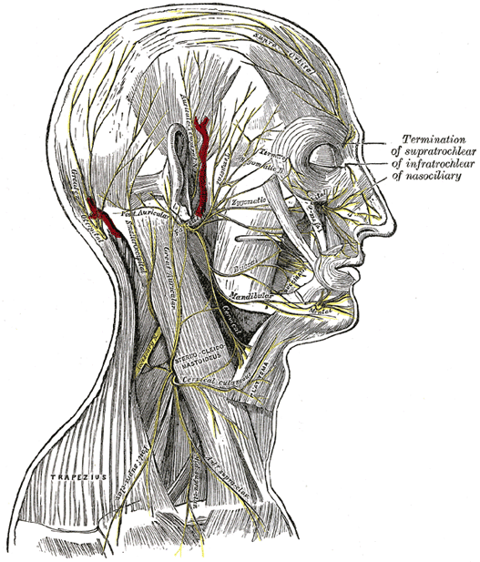
| The Temporal Branches (rami temporales) cross the zygomatic arch to the temporal region, supplying the Auriculares anterior and superior, and joining with the zygomaticotemporal branch of the maxillary, and with the auriculotemporal branch of the mandibular. The more anterior branches supply the Frontalis, the Orbicularis oculi, and the Corrugator, and join the supraorbital and lacrimal branches of the ophthalmic. | |||||||||||||||||||||||||||
| The Zygomatic Branches (rami zygomatici; malar branches) run across the zygomatic bone to the lateral angle of the orbit, where they supply the Orbicularis oculi, and join with filaments from the lacrimal nerve and the zygomaticofacial branch of the maxillary nerve. | |||||||||||||||||||||||||||
| The Buccal Branches (rami buccales; infraorbital branches), of larger size than the rest, pass horizontally forward to be distributed below the orbit and around the mouth. The superficial branches run beneath the skin and above the superficial muscles of the face, which they supply: some are distributed to the Procerus, joining at the medial angle of the orbit with the infratrochlear and nasociliary branches of the ophthalmic. The deep branches pass beneath the Zygomaticus and the Quadratus labii superioris, supplying them and forming an infraorbital plexus with the infraorbital branch of the maxillary nerve. These branches also supply the small muscles of the nose. The lower deep branches supply the Buccinator and Orbicularis oris, and join with filaments of the buccinator branch of the mandibular nerve. | |||||||||||||||||||||||||||
| The Mandibular Branch (ramus marginalis mandibulæ) passes forward beneath the Platysma and Triangularis, supplying the muscles of the lower lip and chin, and communicating with the mental branch of the inferior alveolar nerve. | |||||||||||||||||||||||||||
| The Cervical Branch (ramus colli) runs forward beneath the Platysma, and forms a series of arches across the side of the neck over the suprahyoid region. One branch descends to join the cervical cutaneous nerve from the cervical plexus; others supply the Platysma. | |||||||||||||||||||||||||||
5h. The Acoustic Nerve
(Eighth Nerve)
The acoustic nerve consists of two distinct sets of fibers which differ in their peripheral endings, central connections, functions, and time of medullation. It is soft in texture and devoid of neurilemma. |
|||||
| Cochlear Nerve.—The cochlear nerve or root, the nerve of hearing, arises from bipolar cells in the spiral ganglion of the cochlea, situated near the inner edge of the osseous spiral lamina. The peripheral fibers pass to the organ of Corti. The central ones pass down the modiolus and then through the foramina of the tractus spiralis foraminosus or through the foramen centrale into the lateral or outer end of the internal auditory meatus. The nerve passes along the internal auditory meatus with the vestibular nerve and across the subarachnoid space, just above the flocculus, almost directly medialward toward the inferior peduncle to terminate in the cochlear nucleus. | |||||
| The cochlear nerve is placed lateral to the vestibular root. Its fibers end in two nuclei: one, the accessory nucleus, lies immediately in front of the inferior peduncle; the other, the tuberculum acusticum, somewhat lateral to it. | |||||
| The striæ medullares (striæ acusticæ) are the axons of the cells of the tuberculum acusticum. They pass over the inferior peduncle, and across the rhomboid fossa to the median sulcus. Here they dip into the substance of the pons, to end around the cells of the superior olivary nuclei of both sides. There are, however, other fibers, and these are both direct and crossed, which pass into the lateral lemniscus. The cells of the accessory nucleus give origin to fibers which run transversely in the pons and constitute the trapezium. Of the trapezoid fibers some end around the cells of the superior olivary nucleus or of the trapezoid nucleus of the same or opposite side, while others, crossed or uncrossed, pass directly into the lateral lemniscus. | |||||
| If the further connections of the cochlear nerve of one side, say the left, be considered, it is found that they lie lateral to the main sensory tract, the lemniscus, and are therefore termed the lateral lemniscus. The fibers comprising the left lateral lemniscus arise in the superior olivary and trapezoid nuclei of the same or opposite side, while others are the uninterrupted fibers already alluded to, and these are either crossed or uncrossed, the former being the axons of the cells of the right accessory nucleus or of the cells of the right tuberculum acusticum, while the latter are derived from the cells of the left nuclei. In the upper part of the lateral lemniscus there is a collection of nerve cells, the nucleus of the lateral lemniscus, around the cells of which some of the fibers arborize and from the cells of which axons originate to continue upward the tract of the lateral lemniscus. The ultimate ending of the left lateral lemniscus is partly in the opposite medial geniculate body, and partly in the inferior colliculi. From the cells of these bodies new fibers arise and ascend in the occipital part of the internal capsule to reach the posterior three-fifths of the left superior temporal gyrus and the transverse temporal gyri. | |||||
| Vestibular Nerve.—The vestibular nerve or root, the nerve of equilibration, arises from bipolar cells in the vestibular ganglion, ganglion of Scarpa, which is situated in the upper part of the outer end of the internal auditory meatus. The peripheral fibers divide into three branches: the superior branch passes through the foramina in the area vestibularis superior and ends in the utricle and in the ampullæ of the superior and lateral semicircular ducts; the fibers of the inferior branch traverse the foramina in the area vestibularis inferior and end in the saccule; the posterior branch runs through the foramen singulare and supplies the ampulla of the posterior semicircular duct. |
5i. The Glossopharyngeal Nerve
 |
(N. Glossopharyngeus; Ninth Nerve)
The glossopharyngeal nerve (Figs. 791, 792, 793) contains both motor and sensory fibers, and is distributed, as its name implies, to the tongue and pharynx. It is the nerve of ordinary sensation to the mucous membrane of the pharynx, fauces, and palatine tonsil, and the nerve of taste to the posterior part of the tongue. It is attached by three or four filaments to the upper part of the medulla oblongata, in the groove between the olive and the inferior peduncle. |
| The sensory fibers arise from the cells of the superior and petrous ganglia, which are situated on the trunk of the nerve, and will be presently described. When traced into the medulla, some of the sensory fibers, probably sympathetic afferent, end by arborizing around the cells of the upper part of a nucleus which lies beneath the ala cinerea in the lower part of the rhomboid fossa. Many of the fibers, probably the taste fibers, contribute to form a strand, named the fasciculus solitarius, which descends in the medulla oblongata. Associated with this strand are numerous nerve cells, and around these the fibers of the fasciculus end. The somatic sensory fibers, few in number, are said to join the spinal tract of the trigeminal nerve. |
| The somatic motor fibers spring from the cells of the nucleus ambiguus, which lies some distance from the surface of the rhomboid fossa in the lateral part of the medulla and is continuous below with the anterior gray column of the medulla spinalis. From this nucleus the fibers are first directed backward, and then they bend forward and lateralward to join the fibers of the sensory root. The nucleus ambiguus gives origin to the motor branches of the glossopharyngeal and vagus nerves, and to the cranial part of the accessory nerve. |
| The sympathetic efferent fibers from the nucleus beneath the ala cinerea, the dorsal nucleus, are probably both preganglionic motor fibers and preganglionic secretory fibers of the sympathetic system. The secretory fibers pass to the otic ganglion and from it secondary neurons are distributed to the parotid gland. Some authors describe these fibers as arising from a distinct nucleus the inferior salivatory nucleus, which lies near the dorsal nucleus. |
| From the medulla oblongata, the glossopharyngeal nerve passes lateralward across the flocculus, and leaves the skull through the central part of the jugular foramen, in a separate sheath of the dura mater, lateral to and in front of the vagus and accessory nerves (Fig. 792). In its passage through the jugular foramen, it grooves the lower border of the petrous part of the temporal bone; and, at its exit from the skull, passes forward between the internal jugular vein and internal carotid artery; it descends in front of the latter vessel, and beneath the styloid process and the muscles connected with it, to the lower border of the Stylopharyngeus. It then curves forward, forming an arch on the side of the neck and lying upon the Stylopharyngeus and Constrictor pharyngis medius. Thence it passes under cover of the Hyoglossus, and is finally distributed to the palatine tonsil, the mucous membrane of the fauces and base of the tongue, and the mucous glands of the mouth. |
| In passing through the jugular foramen, the nerve presents two ganglia, the superior and the petrous (Fig. 791). |
| The Superior Ganglion (ganglion superius; jugular ganglion) is situated in the upper part of the groove in which the nerve is lodged during its passage through the jugular foramen. It is very small, and is usually regarded as a detached portion of the petrous ganglion. |
| The Petrous Ganglion (ganglion petrosum; inferior ganglion) is larger than the superior and is situated in a depression in the lower border of the petrous portion of the temporal bone. |
 |
| Branches of Communication.—The glossopharyngeal nerve communicates with the vagus, sympathetic, and facial. |
| The branches to the vagus are two filaments which arise from the petrous ganglion, one passing to the auricular branch, and the other to the jugular ganglion, of the vagus. The petrous ganglion is connected by a filament with the superior cervical ganglion of the sympathetic. The branch of communication with the facial perforates the posterior belly of the Digastricus. It arises from the trunk of the glossopharyngeal below the petrous ganglion, and joins the facial just after the exit of that nerve from the stylomastoid foramen. |
| Branches of Distribution.—The branches of distribution of the glossopharyngeal are: the tympanic, carotid, pharyngeal, muscular, tonsillar, and lingual. |
| The Tympanic Nerve (n. tympanicus; nerve of Jacobson) arises from the petrous ganglion, and ascends to the tympanic cavity through a small canal on the under surface of the petrous portion of the temporal bone on the ridge which separates the carotid canal from the jugular fossa. In the tympanic cavity it divides into branches which form the tympanic plexus and are contained in grooves upon the surface of the promontory. This plexus gives off: (1) the lesser superficial petrosal nerve; (2) a branch to join the greater superficial petrosal nerve; and (3) branches to the tympanic cavity, all of which will be described in connection with the anatomy of the middle ear. |
| The Carotid Branches (n. caroticotympanicus superior and n. caroticotympanicus inferior) descend along the trunk of the internal carotid artery as far as its origin, communicating with the pharyngeal branch of the vagus, and with branches of the sympathetic. |
| The Pharyngeal Branches (rami pharyngei) are three or four filaments which unite, opposite the Constrictor pharyngis medius, with the pharyngeal branches of the vagus and sympathetic, to form the pharyngeal plexus; branches from this plexus perforate the muscular coat of the pharynx and supply its muscles and mucous membrane. |
| The Muscular Branch (ramus stylopharyngeus) is distributed to the Stylopharyngeus. |
| The Tonsillar Branches (rami tonsillares) supply the palatine tonsil, forming around it a plexus from which filaments are distributed to the soft palate and fauces, where they communicate with the palatine nerves. |
 |
| The Lingual Branches (rami linguales) are two in number; one supplies the papillæ vallatæ and the mucous membrane covering the base of the tongue; the other supplies the mucous membrane and follicular glands of the posterior part of the tongue, and communicates with the lingual nerve. |
5j. The Vagus Nerve
(N. Vagus; Tenth Nerve; Pneumogastric Nerve)
The vagus nerve (Figs. 791, 792, 793) is composed of both motor and sensory fibers, and has a more extensive course and distribution than any of the other cranial nerves, since it passes through the neck and thorax to the abdomen. |
|||||||||||||||||||||||
| The vagus is attached by eight or ten filaments to the medulla oblongata in the groove between the olive and the inferior peduncle, below the glossopharyngeal. The sensory fibers arise from the cells of the jugular ganglion and ganglion nodosum of the nerve, and, when traced into the medulla oblongata mostly end by arborizing around the cells of the inferior part of a nucleus which lies beneath the ala cinerea in the lower part of the rhomboid fossa. These are the sympathetic afferent fibers. Some of the sensory fibers of the glossopharyngeal nerve have been seen to end in the upper part of this nucleus. A few of the sensory fibers of the vagus, probably taste fibers, descend in the fasciculus solitarius and end around its cells. The somatic sensory fibers, few in number, from the posterior part of the external auditory meatus and the back of the ear, probably join the spinal tract of the trigeminal as it descends in the medulla. The somatic motor fibers arise from the cells of the nucleus ambiguus, already referred to in connection with the motor root of the glossopharyngeal nerve. | |||||||||||||||||||||||
| The sympathetic efferent fibers, distributed probably as preganglionic fibers to the thoracic and abdominal viscera, i. e., as motor fibers to the bronchial tree, inhibitory fibers to the heart, motor fibers to the esophagus, stomach, small intestine and gall passages, and as secretory fibers to the stomach and pancreas, arise from the dorsal nucleus of the vagus. | |||||||||||||||||||||||
| The filaments of the nerve unite, and form a flat cord, which passes beneath the flocculus to the jugular foramen, through which it leaves the cranium. In emerging through this opening, the vagus is accompanied by and contained in the same sheath of dura mater with the accessory nerve, a septum separating them from the glossopharyngeal which lies in front (Fig. 792). In this situation the vagus presents a well-marked ganglionic enlargement, which is called the jugular ganglion (ganglion of the root); to it the accessory nerve is connected by one or two filaments. After its exit from the jugular foramen the vagus is joined by the cranial portion of the accessory nerve, and enlarges into a second gangliform swelling, called the ganglion nodosum (ganglion of the trunk); through this the fibers of the cranial portion of the accessory pass without interruption, being principally distributed to the pharyngeal and superior laryngeal branches of the vagus, but some of its fibers descend in the trunk of the vagus, to be distributed with the recurrent nerve and probably also with the cardiac nerves. | |||||||||||||||||||||||
| The vagus nerve passes vertically down the neck within the carotid sheath, lying between the internal jugular vein and internal carotid artery as far as the upper border of the thyroid cartilage, and then between the same vein and the common carotid artery to the root of the neck. The further course of the nerve differs on the two sides of the body. | |||||||||||||||||||||||
| On the right side, the nerve passes across the subclavian artery between it and the right innominate vein, and descends by the side of the trachea to the back of the root of the lung, where it spreads out in the posterior pulmonary plexus. From the lower part of this plexus two cords descend on the esophagus, and divide to form, with branches from the opposite nerve, the esophageal plexus. Below, these branches are collected into a single cord, which runs along the back of the esophagus enters the abdomen, and is distributed to the postero-inferior surface of the stomach, joining the left side of the celiac plexus, and sending filaments to the lienal plexus. | |||||||||||||||||||||||
| On the left side, the vagus enters the thorax between the left carotid and subclavian arteries, behind the left innominate vein. It crosses the left side of the arch of the aorta, and descends behind the root of the left lung, forming there the posterior pulmonary plexus. From this it runs along the anterior surface of the esophagus, where it unites with the nerve of the right side in the esophageal plexus, and is continued to the stomach, distributing branches over its anterosuperior surface; some of these extend over the fundus, and others along the lesser curvature. Filaments from these branches enter the lesser omentum, and join the hepatic plexus. | |||||||||||||||||||||||
| The Jugular Ganglion (ganglion jugulare; ganglion of the root) is of a grayish color, spherical in form, about 4 mm. in diameter. | |||||||||||||||||||||||
| Branches of Communication.—This ganglion is connected by several delicate filaments to the cranial portion of the accessory nerve; it also communicates by a twig with the petrous ganglion of the glossopharyngeal, with the facial nerve by means of its auricular branch, and with the sympathetic by means of an ascending filament from the superior cervical ganglion. | |||||||||||||||||||||||
| The Ganglion Nodosum (ganglion of the trunk; inferior ganglion) is cylindrical in form, of a reddish color, and 2.5 cm. in length. Passing through it is the cranial portion of the accessory nerve, which blends with the vagus below the ganglion. | |||||||||||||||||||||||
| Branches of Communication.—This ganglion is connected with the hypoglossal, the superior cervical ganglion of the sympathetic, and the loop between the first and second cervical nerves. | |||||||||||||||||||||||
| Branches of Distribution.—The branches of distribution of the vagus are: | |||||||||||||||||||||||
| |||||||||||||||||||||||
| The Meningeal Branch (ramus meningeus; dural branch) is a recurrent filament given off from the jugular ganglion; it is distributed to the dura mater in the posterior fossa of the base of the skull. | |||||||||||||||||||||||
| The Auricular Branch (ramus auricularis; nerve of Arnold) arises from the jugular ganglion, and is joined soon after its origin by a filament from the petrous ganglion of the glossopharyngeal; it passes behind the internal jugular vein, and enters the mastoid canaliculus on the lateral wall of the jugular fossa. Traversing the substance of the temporal bone, it crosses the facial canal about 4 mm. above the stylomastoid foramen, and here it gives off an ascending branch which joins the facial nerve. The nerve reaches the surface by passing through the tympanomastoid fissure between the mastoid process and the tympanic part of the temporal bone, and divides into two branches: one joins the posterior auricular nerve, the other is distributed to the skin of the back of the auricula and to the posterior part of the external acoustic meatus. | |||||||||||||||||||||||
| The Pharyngeal Branch (ramus pharyngeus), the principal motor nerve of the pharynx, arises from the upper part of the ganglion nodosum, and consists principally of filaments from the cranial portion of the accessory nerve. It passes across the internal carotid artery to the upper border of the Constrictor pharyngis medius, where it divides into numerous filaments, which join with branches from the glossopharyngeal, sympathetic, and external laryngeal to form the pharyngeal plexus. From the plexus, branches are distributed to the muscles and mucous membrane of the pharynx and the muscles of the soft palate, except the Tensor veli palatini. A minute filament descends and joins the hypoglossal nerve as it winds around the occipital artery. | |||||||||||||||||||||||
| The Superior Laryngeal Nerve (n. laryngeus superior) larger than the preceding, arises from the middle of the ganglion nodosum and in its course receives a branch from the superior cervical ganglion of the sympathetic. It descends, by the side of the pharynx, behind the internal carotid artery, and divides into two branches, external and internal. | |||||||||||||||||||||||
| The external branch (ramus externus), the smaller, descends on the larynx, beneath the Sternothyreoideus, to supply the Cricothyreoideus. It gives branches to the pharyngeal plexus and the Constrictor pharyngis inferior, and communicates with the superior cardiac nerve, behind the common carotid artery. | |||||||||||||||||||||||
| The internal branch (ramus internus) descends to the hyothyroid membrane, pierces it in company with the superior laryngeal artery, and is distributed to the mucous membrane of the larynx. Of these branches some are distributed to the epiglottis, the base of the tongue, and the epiglottic glands; others pass backward, in the aryepiglottic fold, to supply the mucous membrane surrounding the entrance of the larynx, and that lining the cavity of the larynx as low down as the vocal folds. A filament descends beneath the mucous membrane on the inner surface of the thyroid cartilage and joins the recurrent nerve. | |||||||||||||||||||||||
| The Recurrent Nerve (n. recurrens; inferior or recurrent laryngeal nerve) arises, on the right side, in front of the subclavian artery; winds from before backward around that vessel, and ascends obliquely to the side of the trachea behind the common carotid artery, and either in front of or behind the inferior thyroid artery. On the left side, it arises on the left of the arch of the aorta, and winds below the aorta at the point where the ligamentum arteriosum is attached, and then ascends to the side of the trachea. The nerve on either side ascends in the groove between the trachea and esophagus, passes under the lower border of the Constrictor pharyngis inferior, and enters the larynx behind the articulation of the inferior cornu of the thyroid cartilage with the cricoid; it is distributed to all the muscles of the larynx, excepting the Cricothyreoideus. It communicates with the internal branch of the superior laryngeal nerve, and gives off a few filaments to the mucous membrane of the lower part of the larynx. | |||||||||||||||||||||||
| As the recurrent nerve hooks around the subclavian artery or aorta, it gives off several cardiac filaments to the deep part of the cardiac plexus. As it ascends in the neck it gives off branches, more numerous on the left than on the right side, to the mucous membrane and muscular coat of the esophagus; branches to the mucous membrane and muscular fibers of the trachea; and some pharyngeal filaments to the Constrictor pharyngis inferior. | |||||||||||||||||||||||
| The Superior Cardiac Branches (rami cardiaci superiores; cervical cardiac branches), two or three in number, arise from the vagus, at the upper and lower parts of the neck. | |||||||||||||||||||||||
| The upper branches are small, and communicate with the cardiac branches of the sympathetic. They can be traced to the deep part of the cardiac plexus. | |||||||||||||||||||||||
| The lower branch arises at the root of the neck, just above the first rib. That from the right vagus passes in front or by the side of the innominate artery, and proceeds to the deep part of the cardiac plexus; that from the left runs down across the left side of the arch of the aorta, and joins the superficial part of the cardiac plexus. | |||||||||||||||||||||||
| The Inferior Cardiac Branches (rami cardiaci inferiores; thoracic cardiac branches), on the right side, arise from the trunk of the vagus as it lies by the side of the trachea, and from its recurrent nerve; on the left side from the recurrent nerve only; passing inward, they end in the deep part of the cardiac plexus. | |||||||||||||||||||||||
| The Anterior Bronchial Branches (rami bronchiales anteriores; anterior or ventral pulmonary branches), two or three in number, and of small size, are distributed on the anterior surface of the root of the lung. They join with filaments from the sympathetic, and form the anterior pulmonary plexus. | |||||||||||||||||||||||
| The Posterior Bronchial Branches (rami bronchiales posteriores; posterior or dorsal pulmonary branches), more numerous and larger than the anterior, are distributed on the posterior surface of the root of the lung; they are joined by filaments from the third and fourth (sometimes also from the first and second) thoracic ganglia of the sympathetic trunk, and form the posterior pulmonary plexus. Branches from this plexus accompany the ramifications of the bronchi through the substance of the lung. | |||||||||||||||||||||||
| The Esophageal Branches (rami æsophagei) are given off both above and below the bronchial branches; the lower are numerous and larger than the upper. They form, together with the branches from the opposite nerve, the esophageal plexus. From this plexus filaments are distributed to the back of the pericardium. | |||||||||||||||||||||||
| The Gastric Branches (rami gastrici) are distributed to the stomach. The right vagus forms the posterior gastric plexus on the postero-inferior surface of the stomach and the left the anterior gastric plexus on the antero-superior surface. | |||||||||||||||||||||||
| The Celiac Branches (rami cæliaci) are mainly derived from the right vagus: they join the celiac plexus and through it supply branches to the pancreas, spleen, kidneys, suprarenal bodies, and intestine. | |||||||||||||||||||||||
| The Hepatic Branches (rami hepatici) arise from the left vagus: they join the hepatic plexus and through it are conveyed to the liver. http://www.theodora.com/anatomy/embryology_index.html | |||||||||||||||||||||||


The large or more aggressive variation of nasopharyngeal carcinoma finds a route through this foramen to reach the cavernous sinus, clivus, and temporal bone. Read More At https://healthjunta.com/foramen-lacerum/
ReplyDelete